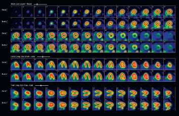
New cardiac imaging technology at the University of Ottawa Heart Institute will allow physicians and researchers to greatly expand their pursuit of advanced heart function studies while ensuring shorter diagnostic wait times for patients.
A new hybrid machine that combines computerized tomography (CT) and positron emission tomography (PET) is expected to reduce wait times to three weeks or less for patients sent for early diagnosis of cardiac conditions that could lead to stroke or heart attack.
The combination of CT and PET blends precise imaging with diagnostic capability more characteristic of invasive angiography, where a catheter is threaded through the blood vessels to the heart to check for clogged arteries.
“We will be focusing more on the population that will most benefit from an advanced technology that has a proven track record as a reliable diagnostic tool,” said cardiologist Dr. Benjamin Chow, Director of Cardiac Radiology at the Heart Institute.
These are often patients who do not necessarily show symptoms but whose physicians suspect a heart condition. CT, for example, uses a special digital camera that can peer inside the vessel wall to show possible calcium buildup. Vessel walls laden with calcium or plaque can narrow to the point of diminishing blood flow to the heart—a condition known as atherosclerosis. Plaque can also rupture or break off, resulting in a blockage that can cut off blood flow altogether, causing a heart attack.
“We can further develop our expertise in all imaging modalities and calculations, which means being able to predict disease in patients where other tests might look normal,” said Dr. Chow.

The Heart Institute is the site of Canada’s National Cardiac PET Centre, the only facility with advanced technologies focused exclusively on cardiac imaging. More than 6,000 patients, including many who are sent from across the country, come through the Institute doors for cardiac imaging tests.
Wait times have grown due to the increasing demand for PET imaging. “This second scanner will now allow us to shorten our wait times and enable more patients to benefit from this increased accuracy,” said cardiologist Dr. Rob Beanlands, Chief of Cardiac Imaging and Director of the PET Centre.
Cardiac imaging at the Heart Institute is also a significant research tool in continued studies to assess viability of the heart and blood flow. The Institute is recognized internationally for its work in assessing and extending the capabilities of PET imaging. Researchers can now detect, for example, even the most subtle changes in how smoothly and rapidly blood flows to the heart after a high-fat meal is eaten.
“We are currently active clinically and in research. This increases our capacity to provide both services, which will help to meet our increasing demand because of the high accuracy of PET,” added Dr. Beanlands.
The Heart Institute is recognized internationally for its work in assessing and extending the capabilities of PET imaging.
In addition to PET’s imaging accuracy, the technology can employ medical isotopes that offer alternatives to the widely used technetium-99. A Heart Institute physics team has been developing such isotopes in response to the critical technetium-99 shortage that resulted from the shutdown of the Chalk River reactor last year. Chalk River is the primary source for the worldwide supply of that isotope. These alternatives offer great promise for reducing heavy reliance on a single imaging substance.
Heart Institute researchers received $2.17 million through the Canadian Institutes of Health Research and the Natural Sciences and Engineering Research Council of Canada, as well as $2 million in federal funds, to develop alternative radioisotope imaging tracers, test them in diagnosing heart disease, and fast-track production and distribution across Canada.
One of these isotopes, rubidium-82 (Rb-82), is produced at the Heart Institute in a small generator about the size of a miniature refrigerator and has been used on site for more than 10 years in patients undergoing PET scans to diagnose coronary artery disease.
Robert deKemp, PhD, is Chief Medical Physicist for Cardiac Imaging. Any patient might wonder how medical physics in the realm of cardiovascular care relates to his or her treatment.
“Our goal and our leadership in this area derives from our ability to quickly and directly move our research techniques from the laboratory into patient care and how doing so can prompt medical intervention earlier than might otherwise happen,” he explained.
DeKemp and his research team have made significant improvements to the isotope generator, which can operate now for eight continuous weeks—longer than the four to six weeks of similar Rb-82 generators.
His work has led to the development of new software to track blood flow in small vessels and at very low volumes. The ability of researchers to investigate tiny vessels permits examination of the microcirculatory effects of diabetes, for example, and the effects of hormones on the heart.
The Heart Institute is one of a handful of cardiac facilities around the world developing advanced imaging techniques to study blood flow and assess risk for coronary artery disease.
Adding a second scanner, deKemp said, “means an ability to do more scans, which creates improved efficiency and accuracy and which can translate into greater cost-effectiveness. But our work here is more concerned with doing the right test for the patient at the right time. That means cardiac research that will translate directly into better treatment options for patients.”

