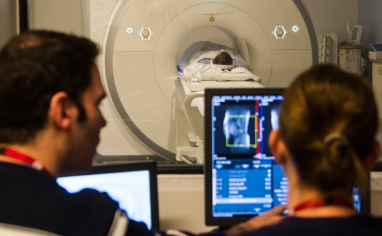
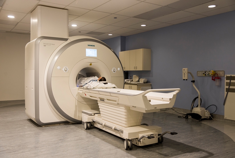
In October, a crane carefully lowered the University of Ottawa Heart Institute’s first dedicated magnetic resonance imaging (MRI) scanner into a narrow access way so that it could be rolled through the exterior wall of the building and into position. By early December, the new MRI facility was fully commissioned and open to serve the 1.2 million residents of the Champlain region.
“We now have in one place all of the technologies needed to image the cardiovascular system and to continue to be a leader in research, education and clinical care,” said cardiologist Alexander Dick, MD, who will co-lead the Heart Institute’s Cardiac MRI program with cardiac radiologist Carole Dennie, MD.
Until now, the Heart Institute had shared The Ottawa Hospital’s MRI facility. But with so many other specialties—including neurology, orthopedics and oncology—clamouring for time on that machine, the wait lists were long and growing, explained Dr. Dick. “Cardiovascular problems often can’t wait for a long time. They need to be imaged quickly. So we needed our own dedicated cardiovascular scanner.”
Not every patient referred to cardiac imaging requires an MRI scan; in fact, most do not, explained Dr. Dick. For simply imaging blockages in the coronary arteries, computed tomography (CT) remains the test of choice, while nuclear imaging can clearly visualize blood flow to the heart muscle.
“But for measuring heart function, heart size and heart shape, cardiac MRI is the current imaging gold standard,” said Girish Dwivedi, MD, PhD, a cardiologist who will lead the MRI facility’s research program. This means that for patients with diseases like cardiomyopathy or congenital heart malformations, an MRI scan will be invaluable for obtaining a clear diagnosis. MRI can also be used to resolve conflicting or inconclusive results obtained from other imaging tests.
Unlike CT, which is a sophisticated three-dimensional X-ray scan, and unlike nuclear imaging techniques, such as a positron emission tomography (PET) or single-photon emission computed tomography (SPECT) scan, MRI uses no radiation. Instead, a patient in the MRI scanner is briefly exposed to a magnetic field. Different types of tissues (muscle, bone, fat, etc.) respond differently at the atomic level to this magnetic exposure. A detector within the MRI scanner converts these responses into an image that shows the different types of tissue in great detail, as well as changes within those tissues.
“With CT or nuclear imaging, you can see that there might be something that’s abnormal in the heart, but you won’t know what that is. MRI can tell you if it’s fat, if it’s clot, if it’s muscle, if it’s tumour. It can give a lot more information than other imaging techniques, at a very, very high resolution, without any radiation and often without any contrast agents,” said Dr. Dick. “Contrast” refers to the injectable dyes required for some types of CT scans, which can cause adverse reactions in some patients.
The clarity and reproducibility provided by MRI also makes it the gold standard for cardiac clinical trials, explained Dr. Dwivedi. “Because cardiac MRI is so accurate, we can reduce the number of patients needed to obtain a meaningful conclusion. So it reduces research costs in the long run because we can both recruit fewer participants and get more accurate data.” Having a dedicated cardiac MRI, added Dr. Dick, “will allow us to participate in a lot more research where MRI is the main imaging end point for the study.”
Because an MRI scan uses significant imaging resources—the team expects to be able to perform around 10 to 15 scans a day—research staff want to make the most of each scan. “Suitable patients undergoing cardiac MRI will be invited to take part in ongoing research studies, which in some cases may mean just enrolling them in a registry,” said Dr. Dwivedi. Such a registry will allow the group to go back and examine correlations between patient outcomes and cardiac abnormalities seen on MRI. In addition, about 20 per cent of the scanner time available will eventually be dedicated to the Heart Institute’s laboratory researchers.
The scanner will also augment the Heart Institute’s educational efforts, said Benjamin Chow, MD, Director of Cardiac Imaging. “Within cardiac imaging, we’re very much focused on training the clinical experts of tomorrow. We have excellent programs in imaging. But the missing link from an educational standpoint has been the MRI component. So this now opens up another avenue of training, thereby attracting individuals from around the world to come and work with us.”
The University of Ottawa Heart Institute Foundation raised the funds over several years to purchase the machine and build the new facility, and the Heart Institute obtained additional funding from the Ontario Ministry of Health and Long-Term Care for ongoing operational costs.
Cardiac MRI remains an expensive test but can be cost-effective when used to minimize the overall amount of testing a patient undergoes. “We don’t want to perform a test, find it inconclusive and then have to order another test. This is inconvenient to patients, increases the cost of health care and may expose patients to additional risk,” said Dr. Chow.
“We’re extremely excited to have this technology and that our patients will have access to it. Our ultimate goal,” he said, “is to provide the best care, and having access to all the latest equipment allows us to do that.”