
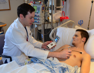
A device about the size of a smartphone is enabling cardiologists to generate images of patients’ hearts at the point of care, enabling them to make more informed diagnoses and even intervene earlier. The result? Improved care and outcomes, and possibly even reduced health care costs.
The device is a portable ultrasound machine with a cardiac probe. It’s called a pocket echo (echo is short for “echocardiogram”). Cardiology fellow Ben Hibbert, MD, PhD, spearheaded its evaluation at the University of Ottawa Heart Institute in 2013, when he was Chief Resident. “It is a transformational development,” he said. In 10 years, he expects every cardiologist—and maybe even every family physician—will have one of these devices in his or her pocket.
Adding Visuals to the Mix
For decades, doctors have used the stethoscope to listen to the heart and deduce what was wrong based on what they heard. It is an important tool, but, said Michel Le May, MD, Director of the Heart Institute’s Cardiac Intensive Care Unit, “There is an element of imprecision.”
“This is the equivalent of an iPhone in our pocket.”
– Michel Le May, MD
Director, Cardiac Intensive Care Unit, UOHI
Then about 40 years ago, there was a giant step forward. With the development of ultrasound, physicians had a non-invasive way to actually see and take pictures of what was happening in the heart and could base their diagnosis and treatment decisions on those pictures. Called echocardiograms, these images were a huge advance in caring for cardiac patients, allowing cardiologists to “see things you can’t see with your ears,” said Dr. Le May.
Echocardiography gives doctors important information about the structure and function of the heart, but the machines are large and bulky, and not always practical in an emergency situation.
Today, physicians at the Heart Institute are using the pocket echo to get instant pictures of a patient’s heart at the point of care.
“This is the equivalent of an iPhone in our pocket,” said Dr. Le May. “The image quality is better than the large machines we had when I was a resident.”
The Impact of Real-time Imaging
In roughly 80 per cent of cases, the information a physician gets from a stethoscope is sufficient. But about 20 per cent of the time, Dr. Le May said, the pocket echo offers new information that changes a diagnosis, informs a treatment plan and even guides an intervention, like the insertion of a needle to drain fluid from a chamber of the heart.
In many of these cases, Dr. Hibbert noted, without the portable echo, an echocardiogram would not be given the same day, or may not be ordered at all. Indeed, in the case of an emergency, there’s simply no time to take a patient to the standard echocardiography machine. And in that emergency situation, the portable echo can help diagnose the problem and aid in carrying out an intervention that can stabilize a patient before surgery.
The pocket echo is not going to replace formal echocardiography, both Dr. Le May and Dr. Hibbert agreed. The portable echo does not have the functionality of the full echocardiograph and its images aren’t as clear. What it can do, however, is screen patients to identify those who don’t need the full echo, reducing unnecessary imaging. And while they haven’t formally studied this aspect of its use, Dr. Hibbert noted, it is likely that the pocket echo will help ensure better use of scarce resources, thereby saving money, without affecting patient care or outcomes.
Part of Everyday Practice
The Heart Institute bought its pocket echo, a General Electric product called Vscan, in 2012, using donated funds. Drs. Le May and Hibbert then set about having cardiology residents routinely use the device on their patients to understand how best it could be used in regular practice. They now have more than 5,000 images from more than 1,000 patients. Dr. Hibbert is working through the data to determine the impact of the pocket echo on patient care and outcomes.
“Probably no one in the country has the kind of data we have,” said Dr. Le May. Drs. Le May and Hibbert have already published articles based on this data and anticipate many more in the future.
Dr. Le May is excited about the potential of the pocket echo. At the Heart Institute, he anticipates that in a few years, every resident and every cardiologist will have one. And it has application well beyond cardiology.
“It is valuable in the intensive care unit, in the emergency room. It’s a great teaching tool,” he said. “In 10 years, it will be the standard of practice, maybe even in family physician's offices.”
For his part, Dr. Hibbert has assimilated the pocket echo into his everyday practice. “When I go and practice elsewhere and don’t have access to it,” said Dr. Hibbert, “I almost feel naked without it. It’s become so much a part of what I do.”