Purpose
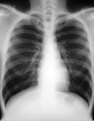
A chest X-ray shows details of the heart, lungs, blood vessels and bones. The radiation created by the X-ray machine passes though the chest and an image is captured on a special digital detector. Different parts of the body absorb different amounts of radiation. The parts of the body that absorb the most radiation, like bones, appear white on the X-ray image. Parts of the body that absorb less radiation, like the heart, will look various shades of grey. Air spaces, like the lungs, look black. A chest X-ray can be used to evaluate how large the heart is and whether there is fluid in the lungs (pulmonary edema) caused by heart failure. Often, more detailed images are required to confirm problems seen on a chest X-ray.
Description
- You will be asked to change into a hospital gown and remove any jewellery in the chest area. This reduces the risk of any interference that can be caused by clothing or jewellery.
- You will be asked to stand in front of the detector in the X-ray room. A medical radiation technologist will confirm that you are positioned correctly.
- The technologist will take two images of your chest: one with you facing the detector and one with you facing the side. You will be asked to take a breath in for each X-ray.
- The images of your chest will be looked at by a radiologist (a doctor who specializes in reading medical images). A report will be sent to the doctor(s) involved in your care.
Patient instructions
- There is no specific preparation required for a chest X-ray. You can eat and drink as usual prior to the test. Take your usual medications unless otherwise directed by your physician.
- There are no restrictions after the chest X-ray.
Additional info
- When you come to the Heart Institute, please check in with central registration in the front lobby. Then proceed to the S-Level and check in at the reception desk.
- A chest X-ray usually takes a few minutes to complete.
- If you have any questions prior to your chest X-ray, please call 613-696-7066, Monday to Friday, 7:00 a.m. to 5:00 p.m.
The image below is interactive
Touring instructions: Click and drag your mouse anywhere in the picture to move about the room. On mobile devices, drag your finger to pan the scene and pinch to zoom in or out. By clicking or tapping the lowercase "i" hotspots, you will learn about the equipment in the room and how it is used.